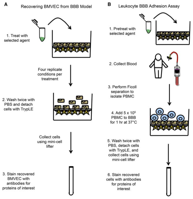
Figure 1.
Schematic representation of BMVEC recovery and leukocyte adhesion assays. The transwell model of the human BBB comprised of BMVEC and astrocytes cocultured on filters with 3-μm pores is established 3 days prior to the commencement of the experiment. (A) The apical side of the BBB transwell may then be treated with the selected agent of interest in four replicate conditions for the desired period of time. Following treatment, the BBB is rinsed twice with PBS, the supernatant discarded, TrypLE added, and the cocultures incubated for 13 min at 37 °C, 5% CO2. The BMVEC are collected by pipetting and gentle scraping with a mini cell lifter and the cells transferred to an appropriate tube for FACS staining. (B) The BBB model is treated for the desired period of time prior to beginning the adhesion assay. PBMC are isolated from blood by density centrifugation with Ficoll-Paque PLUS and the cells (5 × 10⁵) added to the transwell insert for 1 h at 37 °C, 5% CO2. Following incubation, the nonadherent cells are removed by gently washing with PBS twice. The adherent PBMC are collected along with the BMVEC to which they were attached using TrypLE and gentle scraping with the mini cell lifter. The cells are then stained with antibodies for the proteins of interest. [Color figure can be viewed in the online issue, which is available at wileyonlinelibrary.com.]

