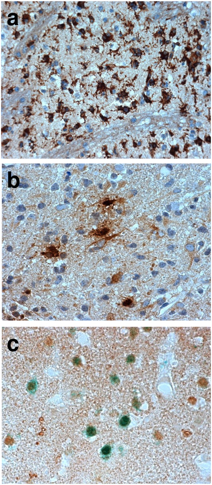
Fig 2. Immunohistochemistry in the mouse brain after temporal middle cerebral artery occlusion (tMCAO).

Macrophage/microglia infiltration (anti-MAC3, brown, a) and lipocalin-2 (LCN2) expression in cells with macrophage/microglia and astrocyte morphology (anti-LCN2, brown, b) in chronic ischemic lesions. LCN2 immunoreactivity is also found in neurons in peri-infarct areas at day 7 after tMCAO (anti-LCN2, brown; anti-NeuN, green; c).
