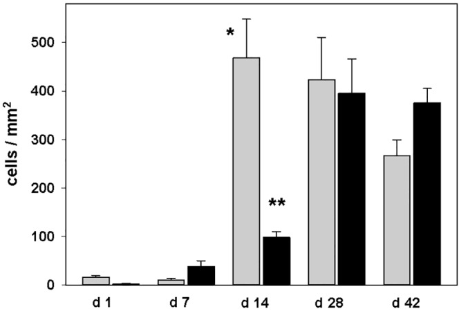
Fig 3. Quantification of lipocalin-2 positive cells (grey) and nonheme iron staining cells (black) in the ischemic striatum in mice after temporal middle cerebral artery occlusion (tMCAO).

Data are presented as mean and standard error; *p<0.001 day 14 vs. days 1 and 7; **p<0.01 day 14 vs. day 7, p<0.001 day 14 vs. days 1, 28 and 42.
