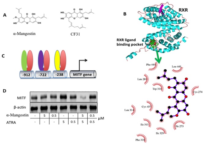
Fig 4. α-Mangostin modulates MITF expression via RXR binding.

A. Chemical structure of α-Mangostin resembles CF31, which targets RXR. B, Molecular docking shows that α-Mangostin is accommodated by the RXR ligand binding pocket. In the top panel RXR is cyan and α-Mangostin is shown as spheres. A close-up view of interactions between RXR and α-Mangostin is shown in the bottom. C. Structure of MITF promoter region. The binding sites of VDR/RXR, RAR/RXR D5 and NURR1/RXR are located at position of -912, -722, and -238 respectively. D. ATRA, a RAR activator, significantly induces expression of MIFT in a dose dependent manner but α-Mangostin blocks expression.
