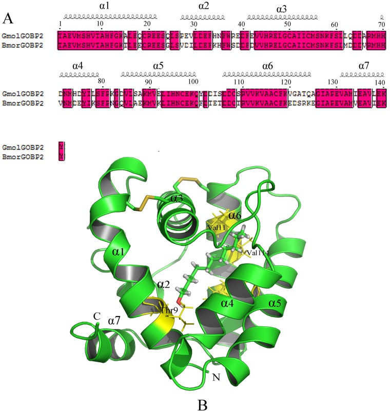
Fig 7. Modeled 3D structure and molecular docking experiments of GmolGOBP2.
(A) Sequence alignment of GmolGOBP2 and BmorGOBP2. α-helices are displayed as squiggles. Strictly identical residues are framed and highlighted with a red background. (B) Overall structure of the GmolGOBP2 and the docking result. Three disulfide bridges are colored in yellow. N-terminal, C-terminal and α-helices are labeled. The top three potential key residues, Thr9, Val111 and Val114, are labeled in black font. Dodecanol is shown as a stick model with the hydroxyl oxygen in red.

