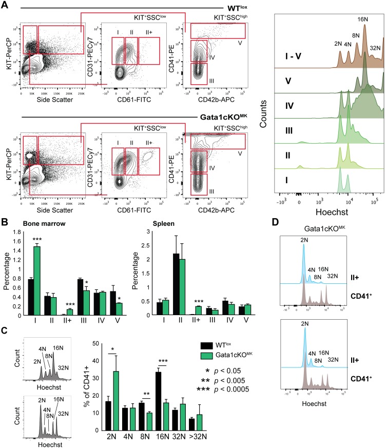
Fig 4. Gata1cKOMKmice have a defect in megakaryopoiesis.
(a) Gating strategy to identify megakaryocytes at consecutive stages of differentiation in the bone marrow and the spleen based on surface marker expression CD31, CD61, CD41 and CD42b. The dot plot depicts the extra population, named II+ found exclusively in Gata1cKOMK bone marrow. On the right we show the ploidy status of the individual subpopulations, thereby justifying our gating strategy. (b) Percentage of megakaryocytes at consecutive stages of differentiation (I-V) of nucleated bone marrow and spleen cells. (c) Ploidy staining of CD41-positive bone marrow and spleen megakaryocytes. The right bar graph depicts ploidy staining of CD41+ in bone marrow. (d) Ploidy status of gated II+ megakaryocyte differentiation stage vs total CD41+ cells in two representative Gata1cKOMK bone marrow samples.

