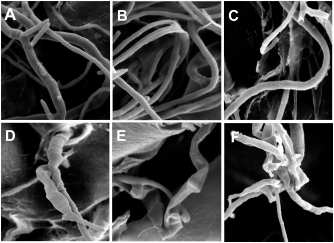
Fig 2. Mycelial morphology of the three tested pathogenic fungi under the electron microscope.
Mycelial morphology of the three tested pathogenic fungi from SE-III-treated medium under the electron microscope. A, B, and C represent the normal condition of M. fructicola, F. oxysporum, and B. dothidea, respectively. D, E, and F represent the hypha of M. fructicola (D), F. oxysporum (E), and B. dothidea (F) from the treated medium.

