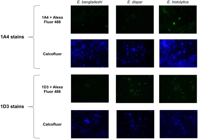
Fig 3. Representative photos from an anti-Jacob2 immunofluorescence microscopy assay.
Isolates were doubled stained with 0.1% Calcofluor White M2R and primary anti-Jacob2 monoclonal antibody (1A4 or 1D3) with goat anti-mouse Alexa Fluor 488. The three species examined were pathogen Entamoeba histolytica (1st column) and commensals Entamoeba dispar and Entamoeba bangladeshi (2nd and 3rd columns). Calcofluor was utilized to identify chitinaceous Entamoeba cysts (2nd and 4th rows).

