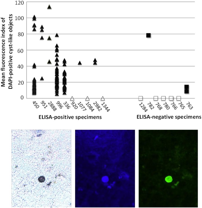
Fig 5. Immunofluorescence staining and image analysis of stool specimens stained with antibody 1A4.
ELISA-positive (triangle) and ELISA-negative (square) specimens were stained with DAPI and antibody 1A4 as described in the text. Eight specimens (open symbols) exhibited no detectable cyst-like objects under the DAPI filter. The bottom panels show a typical cyst detectable by (left to right) bright field, DAPI staining, and antibody 1A4 staining microscopy conducted on a fixed stool slide.

