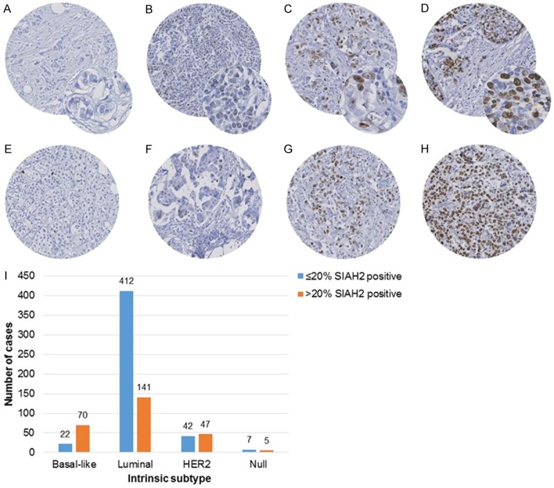
Figure 1.

SIAH2 staining patterns and intrinsic subtypes. A to D exemplify the categories for staining intensity, whereas figures E to H those for proportion of SIAH2-positive tumor cells. A. Negative for SIAH2; B. Weak intensity; C. Moderate intensity; D. Strong intensity. E. Less than 5% of the tumor cells positive for SIAH2, strong staining. F. Moderate staining in 11-20% of the tumor cells. G. Moderate staining in 31-40% of the tumor cells. H. 100% of the tumor cells are strongly stained for SIAH2. I. Shows SIAH2 protein expression within the intrinsic subtypes. Analysis was performed on 746 tumors of the PBC-cohort. Protein expression of SIAH2 was dichotomized on the proportion of positive tumor cells, i.e. ≤ 20% SIAH2-positive versus > 20% SIAH-positive. The intrinsic subtypes were defined by ER, HER2/neu, EGFR and Cytokeratin 5 and classified according to Chan et al. [9] as luminal (positive for ER, negative for HER2/neu), HER2 (positive for HER2/neu), basal (positive for EGFR and/or Cytokeratin 5, negative for ER and HER2/neu) and null (negative for all). SIAH2-positive tumors are especially observed in the basal subtype (76%), in contrast to the luminal subtype (25%).
