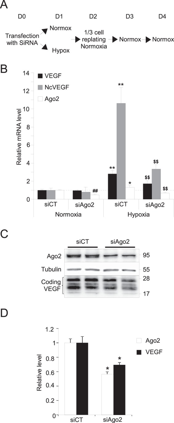
FIG 6.

Ago2 activity is required for the maintenance of VEGF expression after hypoxia. (A) Schema of the experimental design to generate the cDNAs and the proteins analyzed in quantitative PCR (B) and Western blotting (C and D). Briefly, PC3 cells transfected at D0 with either control siRNAs (siCT) or siRNAs targeting Ago2 (siAgo2) were subjected to either normoxia or hypoxia for 24 h (D1) and then replated and placed in normoxia during 72 h (D2 to D4) (see Materials and Methods). (B) cDNA were used to perform qPCR. The results are reported on a histogram as the means ± the SD of two independent experiments, with each sample run in quadruplicate. 36B4 was used to normalize qPCR experiments. *, P ≤ 0.05; **, ##, and $$ all indicate P ≤ 0.005. * and ** were used for siCT in normoxia versus hypoxia. For siCT versus siAgo2, we used ## in normoxia and $$ in hypoxia. (C) Representative Western blot of PC3 cell lysates exposed to hypoxia for 24 h and Ago2, VEGF A antibodies. Tubulin was used as a loading control. (D) After normalization and quantification of Western blots, results are reported on a histogram as the means ± the SD of two independent experiments, with each sample run in duplicate. *, P ≤ 0.05.
