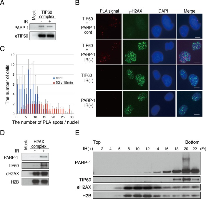
FIG 1.
Functional interaction of PARP-1 and TIP60 histone acetyltransferase. (A) Immunopurified TIP60 complexes were subjected to immunoblot analysis using anti-PARP-1 and anti-TIP60. Nondamaged cells and damaged cells (2 min after 12 Gy of IR) were used for the analysis. (B) PLA analysis using anti-TIP60 and anti-PARP-1 antibodies. The interaction between TIP60 and PARP-1 is detected as PLA spots (red). Costaining with the direct fluorescently labeled anti-γ-H2AX antibody (green) was performed to monitor the DNA damage response. Bars, 10 μm. (C) The numbers of PLA foci within cells were plotted. cont, control. (D) The immunopurified H2AX complex was subjected to immunoblot analyses using anti-PARP-1, anti-TIP60, anti-H2AX, and anti-H2B. Nondamaged cells and damaged cells (2 min after 12 Gy of IR) were used for the analysis. (E) The purified eH2AX complex was subjected to glycerol gradient (10% to 35%) fractionation. Immunoblot analyses using anti-PARP-1, anti-TIP60, anti-H2AX, and anti-H2B were performed. The even-number fractions (Fr) were subjected to Western blotting analysis.

