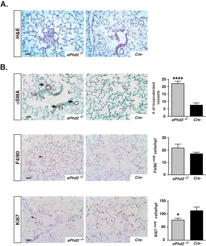
FIG 3.
Histopathological characterization of mice lacking endothelial Phd2. (A) Representative images of H&E-stained lungs from ePhd2−/− mutants and Cre− control mice at 10 weeks of age. (B) Representative images of αSMA-, F4/80-, and Ki67-stained lungs from ePhd2−/− mice and littermate controls. Arrows identify αSMA+ve arterioles (top), F4/80+ve cells (middle), and Ki67+ve cells (bottom). Graphs show quantification of muscularized vessel number/10 HPF, F4/80+ cell number/HPF, and Ki67+ cell number/HPF (n = 5 to 7). Graph bars represent mean values ± SEM. *, P < 0.05; ****, P < 0.0001. Scale bars indicate 50 μm.

