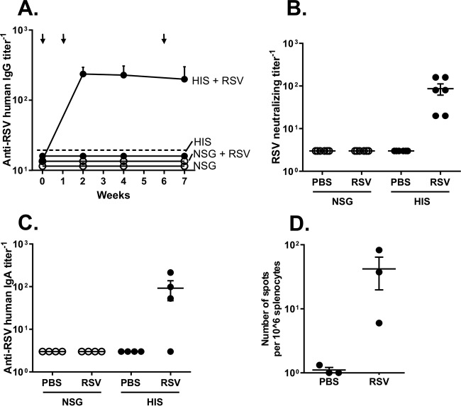
FIG 2.
Adaptive humoral and cellular immune responses in HIS mice following RSV respiratory infection. HIS and NSG mice were infected via the intranasal route with RSV Line 19 (106 PFU/mouse for all mice). (A) Anti-RSV human IgG antibody levels in serum at 0, 2, 4, and 7 weeks analyzed by ELISA. The limit of detection is indicated by the dashed line. The arrows indicate the time points of RSV inoculation. (B) Anti-RSV neutralization titers in mouse serum at 4 weeks postinfection analyzed by virus-neutralizing assay. (C) Anti-RSV human IgA antibody levels in BAL fluid analyzed at 7 weeks analyzed by ELISA. In panels A to C, data are shown as the means ± SEMs for 4 to 6 mice per group. (D) Results of analysis of RSV-specific human IFN-γ-positive spleen cells isolated from RSV-infected HIS and NSG mice at 7 weeks by ELISPOT assay.

