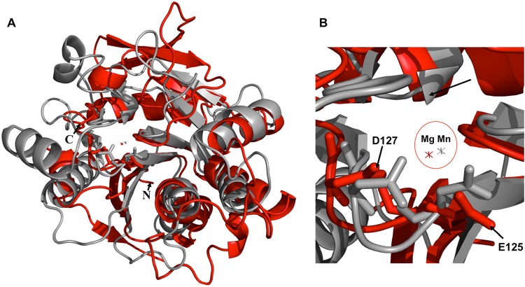
FIG 2.
Comparison of H3 and the PBCV-1 protein A64R. (A) Structural superimposition of H3 (red) and the PBCV-1 protein A64R (gray). (B) Closeup view of the metal coordination site in the superimposition. The Mg2+ ion of H3 (red) is close to the Mn2+ ion of A64R (gray). H3 residues E125 and D127 are the “E/DxD” motif.

