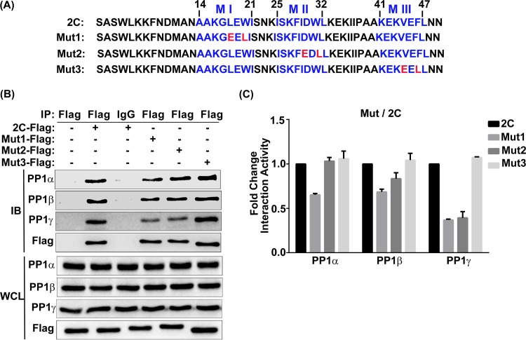
FIG 4.
Interaction of the individual mutated EV71 2C PP1-binding motifs with PP1c. (A) Analysis of the protein sequences of wild-type 2C in comparison with those of 2C mutants Mut1, Mut2, and Mut3. The three PP1-binding motifs of 2C are displayed in blue, and the amino acids replaced by E or L are in red. Numbers indicate the amino acid location in the 2C protein. (B) 293T cells were transfected with a vector, 2C-Flag, or a Flag-tagged mutated 2C sequence. At 24 h posttransfection, cells were harvested and lysed. The whole-cell lysate was immunoprecipitated with anti-Flag or control mouse IgG, and the immunoblots were probed using the corresponding antibodies. (C) Quantification of the proteins in the IP panels from panel B using ImageJ software; the results shown (mean ± SD fold change) are representative of those from three independent experiments.

