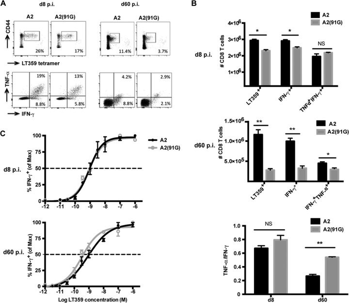
FIG 2.
A2(91G) infection recruits memory CD8 T cells having higher polycytokine functionality and functional avidity. (A) Representative dot plots of splenic DbLT359 tetramer+ CD8 T cells surface stained with anti-CD44 and of intracellular costaining with anti-IFN-γ and anti-TNF-α of anti-CD8 surface-stained spleen cells after LT359 peptide stimulation at days 8 and 60 p.i. (no IFN-γ+ or TNF-α+ cells were detected in the absence of LT359 peptide [data not shown]). (B) Numbers of DbLT359 tetramer+, IFN-γ+, and IFN-γ+ TNF-α+ CD8 T cells at days 8 and 60 p.i. (top and middle) and ratios of TNF-α+ to IFN-γ+ CD8 T cells (bottom). Values represent the means and SEM for three experiments using 3 or 4 mice per group, *, P < 0.05; **, P < 0.005; NS, not significant. (C) LT359 peptide dose-response curves for intracellular IFN-γ staining of splenic CD8 T cells from A2- and A2(91G)-infected mice at day 8 p.i. (top) and day 60 p.i. (bottom). EC50 values were as follows: at day 8 p.i., 9.0 × 10⁻¹⁰ M for A2 and 8.5 × 10⁻¹⁰ M for A2(91G) (P = 0.96); and at day 60 p.i., 8.71 × 10⁻¹⁰ M for A2 and 3.3 × 10⁻¹⁰ M for A2(91G) (P = 0.017). Experiments were repeated twice, with 3 mice per group.

