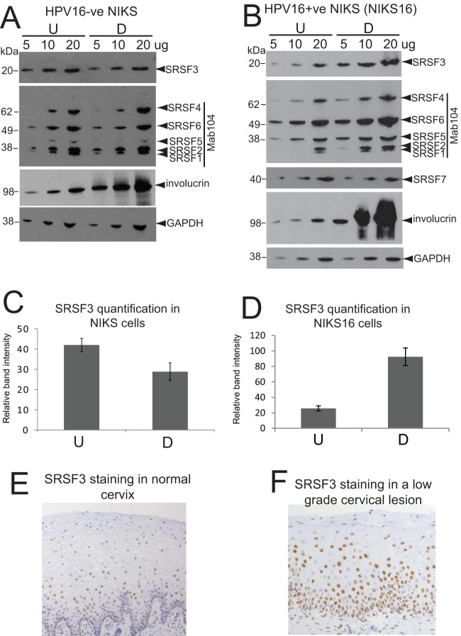
FIG 3.
SRSF3 levels increase upon differentiation of HPV16-infected cells (A) SRSF1, -2, and -3 levels do not alter upon differentiation of HPV-negative NIKS cells. Semiquantitative Western blot analysis of levels of SR proteins in undifferentiated (U) and differentiated (D) NIKS cells. Protein extracts (5, 10, or 20 μg) were loaded as indicated above the blots. SRSF3 was detected with the specific monoclonal antibody 7B4. SRSF1, -2, -4, -5, and -6 were detected with Mab104. SRSF7 was not tested in this experiment because its levels did not change upon HPV infection (24). Involucrin was detected as a control for differentiation of epithelial cells. GAPDH was detected as a protein loading control. (B) SRSF1, -2, and -3 levels are increased in differentiated NIKS16 cells. Semiquantitative Western blot analysis of the levels of SR proteins in undifferentiated (U) and differentiated (D) NIKS16 cells was performed. Protein extracts (5, 10, or 20 μg) were loaded as indicated above the blots. SRSF3 and -7 were detected with specific monoclonal antibodies. SRSF1, -2, -4, -5, and -6 were detected with Mab104. Involucrin was detected as a control for differentiated epithelial cells. GAPDH was detected as a protein loading control. The experiments in panels A and B were carried out at least three times, with very similar results obtained each time. (C) Quantification of SRSF3 levels in undifferentiated (U) and differentiated (D) HPV-negative NIKS cells. The graph shows the means and standard deviations from three separate experiments. Values were calculated relative to the GAPDH levels. (D) Quantification of SRSF3 levels in undifferentiated (U) and differentiated (D) NIKS16 cells. The graph shows the means and standard deviations from five separate experiments. Values were calculated relative to the GAPDH levels. (E) Immunohistochemical staining of a representative normal cervical epithelium (10 normal lesions were stained) with an antibody against SRSF3. Note that the strongly stained nuclei are present mainly in the lower epithelial layers. (F) Immunohistochemical staining of a representative low-grade cervical lesion (10 low-grade lesions were stained) with an antibody against SRSF3. Note the strongly stained nuclei in the middle to upper epithelial layers. The picture in panel E is taken at a lower magnification than that in panel F to show that the staining pattern is consistent over a wide area of tissue.

