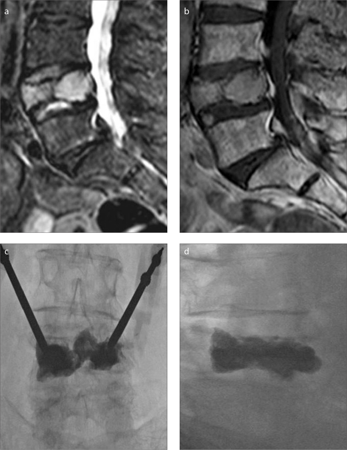
Figure 2. a–d.
A 76-year-old-female patient with multiple myeloma. Grade 3 vertebral height loss on L4 vertebral body is shown as hyperintense signal on STIR sagittal image (a) and isointense signal on sagittal T1-weighted image (b). PV procedure performed on L4 vertebral body with bipedicular approach (c, d).

