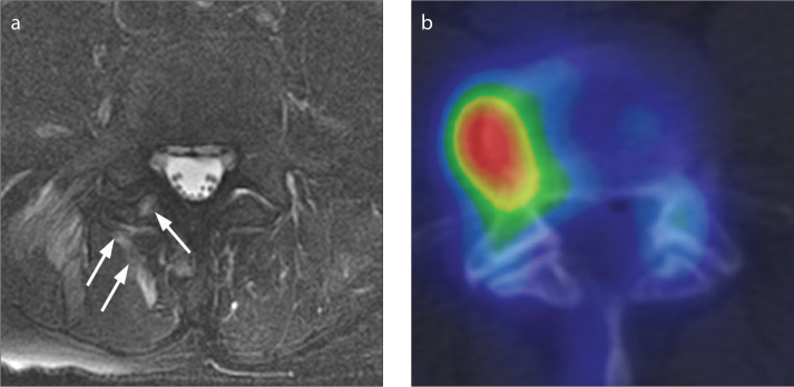
Figure 2. a, b.
MRI and 99mTc MDP SPECT/CT images of a 64-year-old man taken six days apart. The facet joint was rated as abnormal on MRI and normal on 99mTc-MDP SPECT/CT. Axial T2-weighted MRI with fat saturation (a) demonstrates T2 hyperintensity within the inferior articular process of the right L4/L5 facet joint and the surrounding soft tissues (arrows). An axial 99mTc-MDP SPECT/CT (b) image of the lumbar spine demonstrates increased activity within a right lateral osteophyte at L4/L5, but normal activity within the right L4/L5 facet joint itself.

