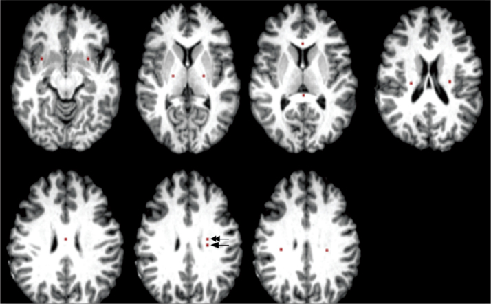
Figure 1.
Placement of regions of interest (ROIs). ROIs of normal appearing white matter (NAWM) were drawn from the genu, splenium and body of the corpus callosum, bilateral corona radiata, superior longitudinal fasciculus, posterior limb of internal capsule, and uncinate fasciculi. Lesion (arrow) and perilesional (double arrow) ROIs were drawn on the left frontal white matter.

