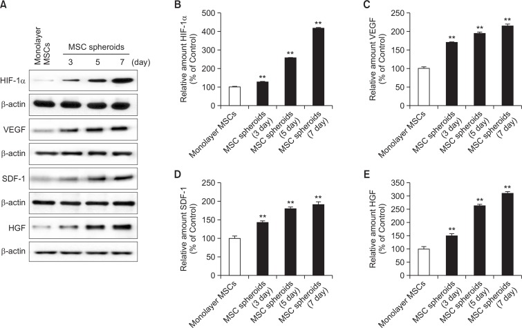
Fig. 2.
Expression of hypoxia-induced angiogenic cytokines in MSC spheroids. (A) Western blot analysis for hypoxic inducible factor-1α (HIF-1α), vascular endothelial growth factor (VEGF), stromal cell derived factor (SDF), and hepatocyte growth factor (HGF) in monolayer MSCs and MSC spheroids cultured for 3, 5, and 7 days. (B-E) Relative expression of HIF-1α, VEGF, SDF, and HGF normalized to that of β-actin. Values represent means ± SEM. **p<0.01 vs. monolayer MSCs.

