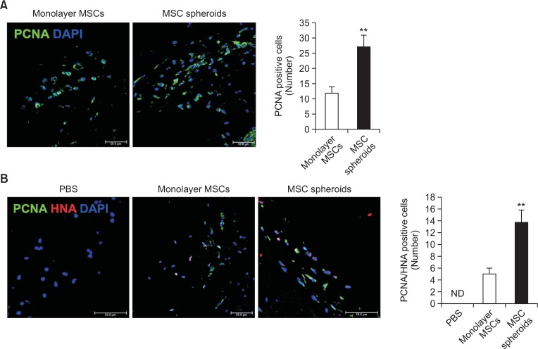
Fig. 5.
Proliferation of MSC spheroids in vivo. (A) Immunofluorescent staining of proliferating cell nuclear antigen (PCNA; green) in murine ischemic limb sites 3 days after transplantation of monolayer MSCs and MSC spheroid cultured for 7 days (scale bar=50 μm). Bar graph shows the number of PCNA-positive cells 3 days after transplantation. Values represent means ± SEM. **p<0.01 vs. monolayer MSCs. (B) Immunofluorescent staining of PCNA (green) and human nucleic antigen (HNA; red) in murine ischemic limb site at 3 days after transplantation of monolayer MSCs and MSC spheroids cultured for 7 days (scale bar=50 μm). Bar graph shows the number of PCNA and HNA double-positive cells 3 days after transplantation. Values represent means ± SEM. **p<0.01 vs. monolayer MSCs.

