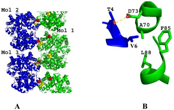
Fig. 1. Structure of DeoxyHb S (PDB code 2HBS).
(A) Ribbon figure of the crystal packing of deoxyHb S. (B) The pathological β2Val6 in one strand (blue) interacts with a hydrophobic pocket formed by β1Ala70, β1Phe85, and β1Leu88 from the β1 subunit of a heterotetramer positioned in the adjacent polymeric strand (green). This interaction is stabilized by a hydrogen-bond contact between β2Thr4 and β1Asp73.

