
Fig. 2. Crystal structure of COHb S.
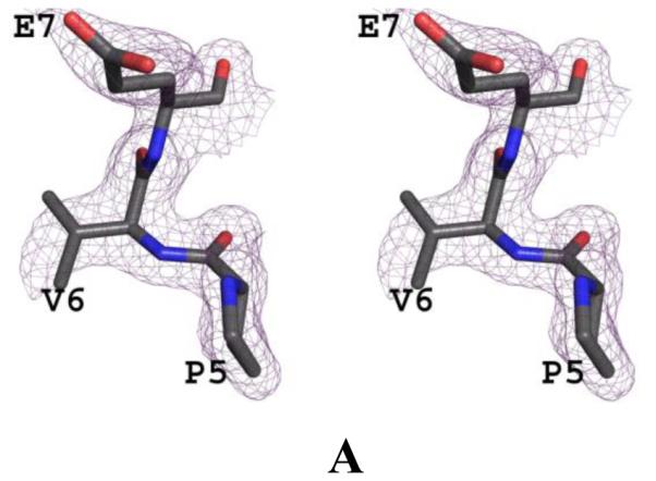
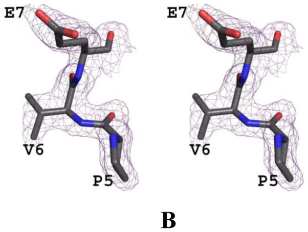
(A) Stereo-view of the initial electron density (2Fo-Fc) map with alanine at the 6th position of the β-subunit during the refinement, contoured at 1.0σ. (B) Stereo-view of the final 2Fo-Fc map with valine at the 6th position of the β-subunit during the refinement, contoured at 1.0σ. The two maps are superimposed with the final refined model.