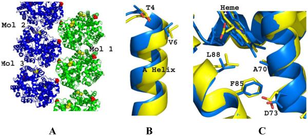
Fig. 3. Structure analysis of COHb S and DeoxyHb S.
(A) Ribbon figure of the crystal packing of COHb S. (B) The β-subunit A helix (with βVal6 and βThr4) of COHb S (yellow) and DeoxyHb S (blue) after superposing the β2-subunits (3-138 residues) of the two structures. (C) The hydrophobic acceptor pocket formed by βAla70, βPhe85, and βLeu88 of COHb S (yellow) and DeoxyHb S (blue) after superposing the β1-subunits (3-138 residues) of the two structures.

