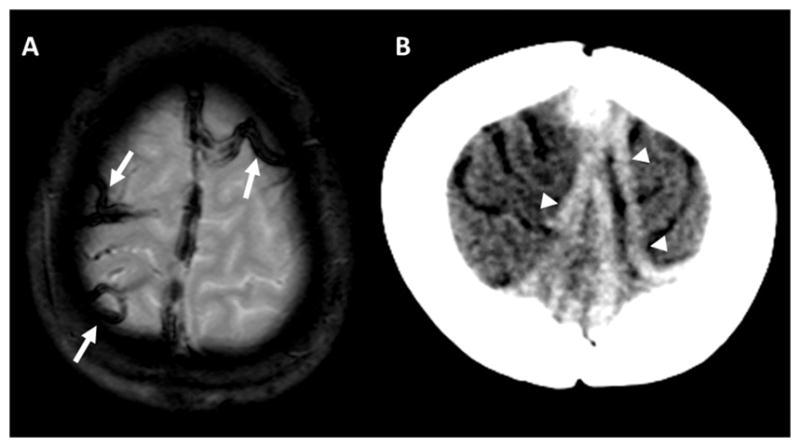
Figure 1.

A) Gradient echo image demonstrates thickened, cord-like areas of hypointensity over the vertex, compatible with thrombosed cortical veins (arrows). B) Non-contrast head CT in a different patient demonstrates abnormal, cord-like densities over the vertex extending to the superior sagittal sinus, also compatible with CVT (arrowheads).
