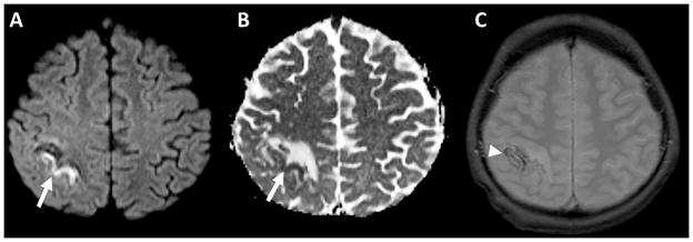
Figure 2.
Diffusion-weighted image A) and apparent diffusion coefficient map B) demonstrates a gyriform focus of restricted diffusion (arrows) surrounded by increased diffusivity, characteristic of acute venous ischemia. C) Gradient echo images in the same patient demonstrate foci of susceptibility compatible with hemorrhage (arrowhead).

