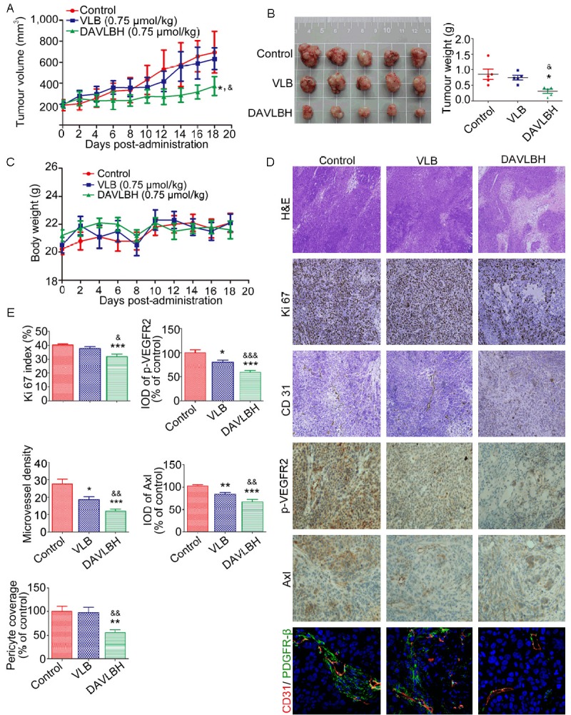
Figure 6.

DAVLBH inhibited tumor growth and tumor angiogenesis in a HeLa xenograft model. A. DAVLBH inhibited HeLa xenograft tumor growth, as measured by tumor volume. HeLa cells (1 × 10⁷ cells per mouse) were injected subcutaneously into 5- to 6-week-old BABL/c (nu/nu) female mice. After the tumors were established (approximately 200 mm3), mice were injected intravenously (i.v.) with saline, VLB or DAVLBH every two days for a total of 9 injections. Differences in tumor volumes on the 18th day were statistically analyzed using a one-way ANOVA, n = 5. *P < 0.05 compared with the control group; &P < 0.05 compared with the VLB-treated group. B. Tumors were removed from the mice and imaged at the end of treatment (left panel, n = 5), and the tumor weights were calculated (right panel, n = 5). C. Mouse body weight curves are shown (n = 5). No significant differences in body weights were observed between the control and the VLB- or DAVLBH-treated groups. D. Representative figures of the H&E staining, immunohistochemical and immunofluorescence analyses of tumors from mice that were sacrificed at the end of the experiment. The H&E staining is at 40× magnification; the immunohistochemical analyses with anti-Ki67 and anti-CD31 are at 100× magnification; the immunohistochemical analyses with anti-Axl and anti-p-VEGFR2 are at 200× magnification; and the immunofluorescence analyses with anti-CD31 (red), anti-PDGFR-β (green) and the nuclei (blue) are at 40× magnification. E. Graphs show the quantitative effect. The immunohistochemical and immunofluorescence results were calculated using Image-Pro Plus 6.0 software. The data are presented as mean ± SEM, n = 5. *P < 0.05, **P < 0.01, and ***P < 0.001 compared with the control group. &P < 0.05, &&P < 0.01 and &&&P < 0.001 compared with the VLB-treated group.
