
Figure 1.
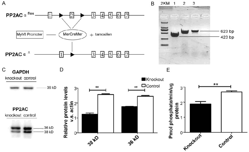
Construction and identification of PP2ACα knockout mice. A. The recombination process and the loxP sites used for PP2ACα knockout. B. Prim PP2A-P5F and PP2A-P5R were used to detect PP2ACα cDNA. The solid line represents detecting PP2ACα cDNA was 200 bp shorter than the control. Quantitative real-time PCR assay was carried out to examine the expression levels of PP2ACα gene. C. Western blot demonstrates PP2AC protein levels (36 and 38 kDa) of mice heart protein extracts from the knockout group and the control group 10 days after tamoxifen injection. D. Statistical analysis for the western blot. E. Phosphatase activity was elevated both in knockout and control groups. n ≥ 6 in each group, *P < 0.05, **P < 0.01.