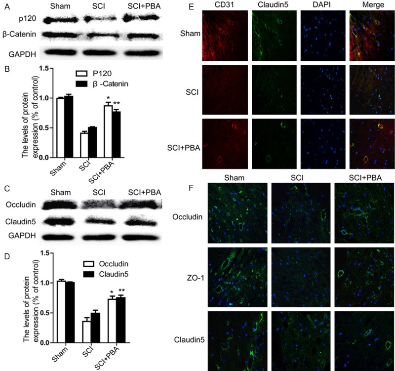
Figure 2.

PBA prevents loss of tight junction and adheren junction proteins after SCI. A. Representative western blots of adherens junction proteins β-catenin, P120 in the sham, SCI model and SCI model treated PBA groups. B. Quantification of western blot data from A. *P < 0.01,**P < 0.01 versus the SCI group, Mean values ± SEM, n = 5. C. Representative western blots of tight junction proteins Occluding, Claudin5 in the sham, SCI model and SCI model treated PBA groups. D. Quantification of western blot data from C. *P < 0.01, **P < 0.01 versus the SCI group, Mean values ± SEM, n = 5. E. Representative micrographs showing (original magnification × 400) double immunofluorescence with Claudin5 (green) and CD31 (endothelial cell marker, red), nuclei are labeled with DAPI (blue) in each group. F. Occludin, Claudin5, and ZO-1 staining (green) results of the sham, SCI group and SCI rat treated with PBA group.
