
Figure 5.
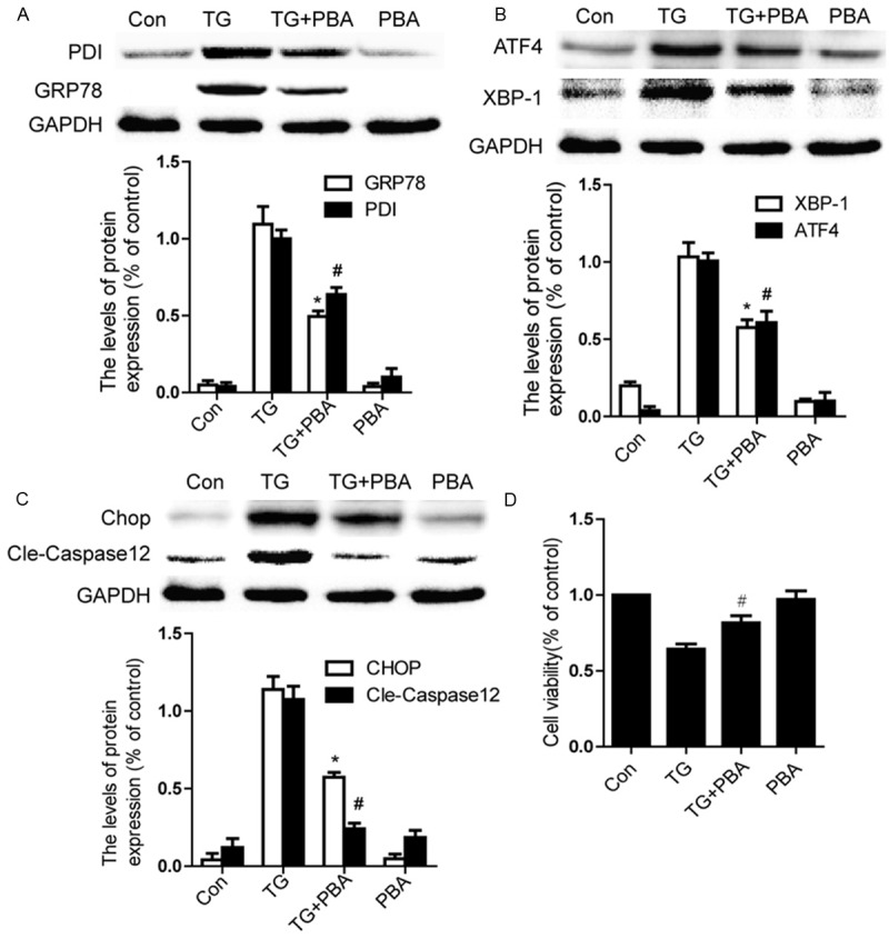
PBA inhibits ER stress and apoptosis in TG-treated endothelial cells. Endothelial cells were treated with TG (10 μM) or together with PBA (1 mM) or PBA (1 mM) alone for 6 h. A. Representative western blots and quantification data of ER stress markers GRP78, PDI in each group cells. B. Representative western blots and quantification data of ER stress markers ATF4 and XBP-1 in each group cells. C. Representative western blots and quantification data of apoptosis-regulating proteins CHOP, Caspase-12, in each group cells. D. MTT results of PBA-treated endothelial cells induced by TG. All experiments were repeated three times. All data represent Mean values ± SEM, *P < 0.01, #P < 0.01 versus TG treated cells.