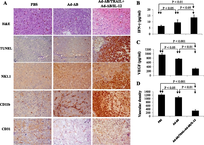
Fig. 5.

Histopathological, immunohistochemical and apoptosis assessments of the harvested tumor tissues from different animal groups. Three days post-treatment of HCC-bearing mice with PBS, Ad-ΔB, or Ad-ΔB/TRAIL+Ad-ΔB/IL-12 (at a dosage regimen of 1 × 10¹⁰ VP in 200 μL PBS of each virus; repeated three times every other day), the mice were euthanized under general anesthesia and their liver tumor tissues were harvested and prepared for histopathological, immunohistochemical (IHC) and apoptosis assessments, and measurement of intra-tumor levels of IFN-γ and VEGF. a Photographs of H&E: hematoxylin and eosin staining of tumor tissues for histopathology; TUNEL: Terminal uridine deoxynucleotidyl transferase dUTP nick end labeling staining assay of apoptotic cells in tumor tissue; NK1.1: IHC staining of infiltrated natural killer cells; CD11b: IHC staining of recruited antigen-presenting cells (dendritic cells and macrophage); and CD31: IHC staining of tumor CD31-positive microvessels endothelial cells. Original magnification: × 400. b and c are quantitative ELISA assays of the intratumor expression levels of IFN-γ and VEGF, respectively, at their protein levels. d The mean microvessel density for each treatment group was determined by counting CD31-positive vessels in 10 high-power fields. Each experiment was performed at least three times, and data shown are from representative experiments. Values of (b), (c) and (d) represent the mean ± SE
