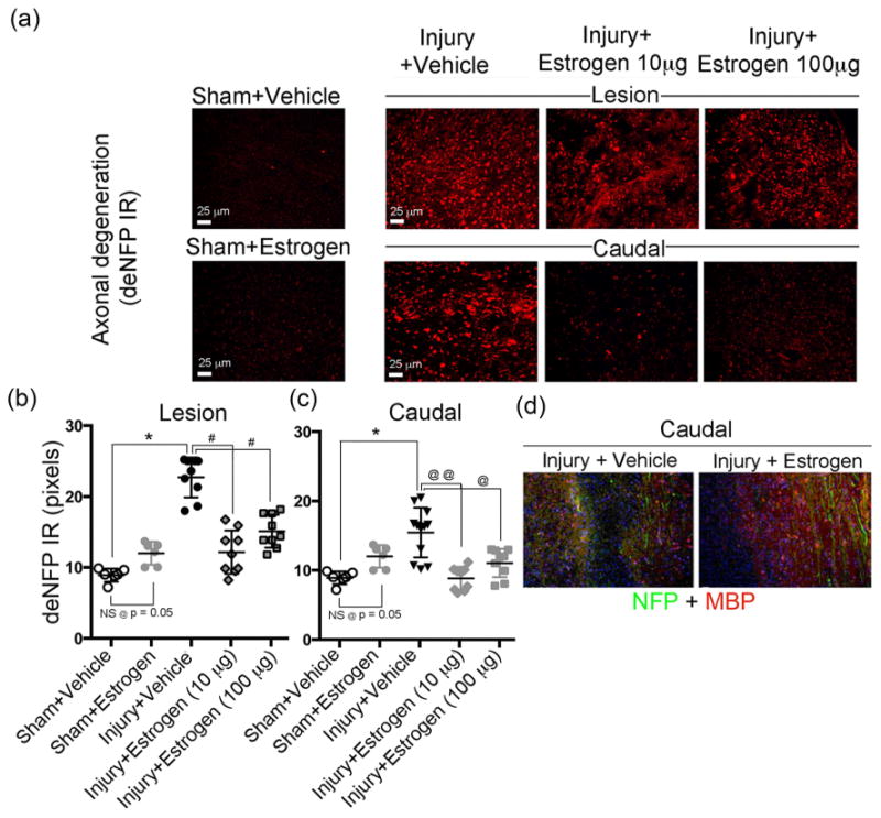
Fig. 3.

Low dose estrogen therapy reduces axonal degeneration and preserves axons following chronic SCI. Thin frozen cross sections (5 μm) were obtained from lesion and caudal penumbra spinal cord tissues (6 weeks post-SCI). Sections were stained with antibody recognizing deNFP. (a) Representative images from the four treatment groups (200× magnification). (b, c) Quantification of deNFP fluorescence for determination of axonal damage. Pixels were quantified using ImageJ. Sham + vehicle and sham + estrogen showed no significant difference at p = 0.05. Significant differences from sham values were indicated by *P < 0.001; compared to injury + vehicle (lesion) as #P < 0.01, and compared to injury + vehicle (caudal penumbra) as @P < 0.05 or @@P < 0.01. (c) Immunofluorescent staining of caudal penumbra of spinal cord tissue from SCI rats following treatment with vehicle or estrogen (10 μg) for nuclei with Hoescht stain (blue), axons with NFP antibody (green), and myelin or myelin forming cells with MBP antibody (red) (100× magnification). n = 3.
