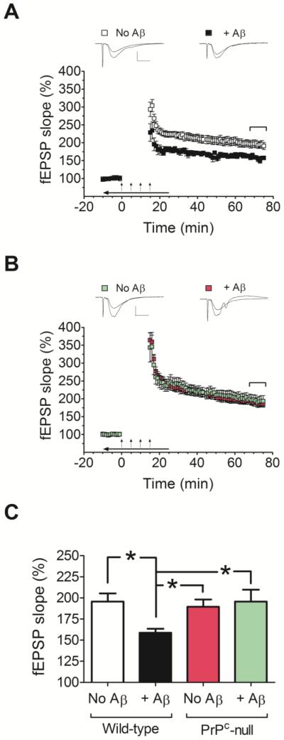
Fig. 3.
Comparison of the effects of Aβ oligomers on LTP in hippocampal slices from wild-type and PrP-null mice. (A and B) Field potentials were recorded from the CA1 region of hippocampal slices from FVB/N wild-type mice (A) or PrPC-null mice (FVB/N background) (B), in the absence or presence of Aβ oligomers (500 nM). (C) Average fEPSP slopes during the last 10 minutes of field potential recordings derived from data shown in panels A and B. Note that hippocampal slices from PrPC null mice perfused with Aβ oligomers did not show impaired LTP. A two-way ANOVA showed a significant interaction between genotype and treatment (Aβ) (F(1, 39)= 4.705, p= 0.036). The oligomers were bath applied for 10 minutes before establishing baseline; during tetanic stimulation (arrows); and for an additional 10 minutes after LTP induction (horizontal arrow bar). Representative traces of individual fEPSPs before and after LTP stimulation are shown on the top of panels A and B (vertical bar: 1 mV; horizontal bar: 10 ms). The mean values are based on 10-11 independent experiments (one slice per animal per treatment), and error bars represent SEM. Statistical significance is expressed as * for p<0.05.

