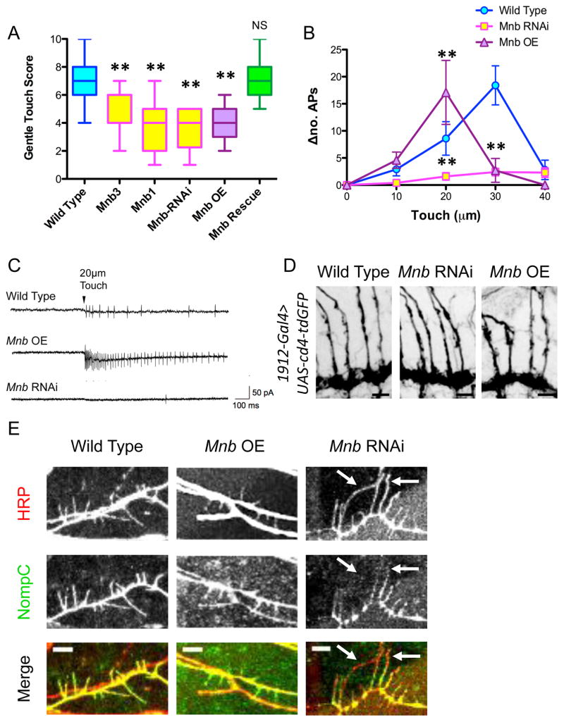
Figure 7. Mnb mutant class III da neurons exhibit a reduced mechanosensitive response due to altered morphology and abnormal localization of NompC.
(A) Quantification of the larval behavioral response to gentle touch reveals defects in the mnb mutant and RNAi animals, as well as in the mnb overexpressing animals (two stars indicate P < 0.0001; for mnb1 rescued with 1912-Gal4>UAS-mnb, P = 1.00; n = 24, 19, 19, 20, 19, and 20 larvae for wild type, mnb3, mnb1, mnb RNAi, and mnb OE, and mnb rescue, respectively). Graph is a box plot with min to max whiskers, which include all datapoints. (B) Extracellular electrophysiological recordings from class III da neurons expressing mnb-RNAi and mnb OE indicate an alteration in the number of action potentials (APs) fired per second for a mechanical stimulus of a particular intensity compared with baseline (Δno. APs) (two stars indicate P < 0.005; n = 8, 9, and 7 larvae for wild type, mnb RNAi, and mnb OE, respectively). (C) Representative recording traces from wild type, mnb OE, and mnb-RNAi neurons for a stimulus of 20-μm intensity. (D) Immunohistochemical staining of larval fillets expressing UAS-cd4-tdGFP specifically in class III da neurons (driven by 1912-Gal4) with antibodies against GFP reveals that the axon terminals of wild type, mnb RNAi, and mnb OE class III da neurons appear similar. Scale bars are 30 μm. (E) Immunohistochemical staining of wild type, mnb OE and mnb RNAi larval fillets with anti-nompC and anti-HRP reveals altered patterns of NompC localization. NompC fills the terminal branches of wild type and mnb OE neurons. In mnb RNAi neurons, NompC is apparent in the proximal regions of the terminal branches, but is not localized throughout the entire terminal branch (white arrows). Scale bars are 5 μm.

