
Figure 6.
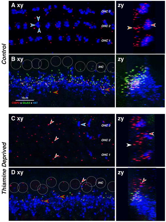
Two weeks of postnatal thiamine deprivation (Post-2) can cause loss of IHC afferent synapses and MOC efferent terminals. Each image is the maximal projection of a confocal z-stack through the IHC (B,D) or OHC (A,C) areas, viewed either in the xy (acquisition) plane (left) or the zy (digitally rotated) plane (right). In normal IHCs, afferent synapses appear as pairs of red (anti-CtBP2) and green (anti-GluA2) puncta (B,D, red-green arrowheads). Positions of IHC nuclei are shown as dotted white circles (B,D). In normal OHCs, presynaptic ribbons are present, but no GluA2 puncta are visible. In thiamine-deprived IHCs, many GluA2 puncta are missing leaving orphan ribbons (D, white-red arrowhead). In thiamine deprived OHCs, the density of ribbons is reduced. OC efferent terminals appear in the blue (anti-VAT) channel. In control OHCs, 2-5 OC terminals contact the base of each OHC (A, white-blue arrowheads). In thiamine-deprived OHCs (C), density of OC terminals is greatly decreased (C). Scale in B applies to all micrographs, which are from the 11 kHz region of 22-wk old cochleas. See text for further description.