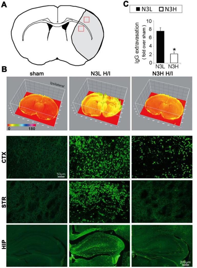
Figure 3.
n-3 PUFAs dramatically reduce extravasation of plasma-derived immunoglobulin G (IgG) at 48 hours after H/I. (A) The regions of interest for IgG measurements in cortex and striatum are marked by red squares. (B) Representative images of IgG expression using a 3D model (top panels) and immunostaining for rat IgG extravasation (lower panels) in cortex (CTX) and striatum (STR) (scale bar=50µm) and in hippocampus (scale bar=200µm) after H/I. (C) Quantification of IgG-positive area as determined by immunohistochemical staining. *p≤0.05 vs N3L H/I, data are mean±s.d., n=6 per group.

