Figure 1.
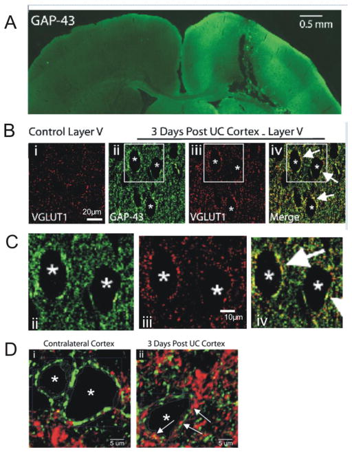
New aberrant excitatory terminals are present in the UC cortex. A. Immunoreactivity (IR) for GAP-43 in lesioned cortex (green). Prominent IR is seen within the undercut and in the ipsilateral cortical and subcortical regions, but not in homotopic regions of uninjured contralateral cortex. Scale bar: 0.5 mm. B. Single plane layer V confocal images. Bi: Diffuse VGLUT1-IR in control cortex (red). Bii-Biii: GAP-43-IR and VGLUT1-IR from dual-reacted section of UC cortex. Perisomatic VGLUT1 puncta are more prominent in UC vs control (Bi vs Biii). Biv: Merged image of Bii and Biii shows some perisomatic close appositions of VGLUT1 and GAP-43 (white arrows). Asterisks mark the same 3 presumptive Pyr cells in Bii-Biv. Scale bar in Bi: 20 μm for Bi-iv. C. Enlarged images of B ii, iii, and iv to show colocalization of GAP43 and VGLUT1. Scale bar in Ciii for Cii-iv. D. Single plane confocal images of layer V pyramidal neurons (asterisks) in contralateral (i) and UC (ii) cortex processed for VGAT1 (green) and VGLUT1 (red) immunoreactivity. Arrows in Dii: examples of VGLUT1 positive puncta in the perisomatic region of a layer V Pyr neuron (asterisk). Scale bar in D: 5 μm.

