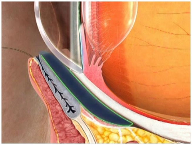
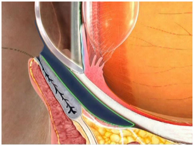
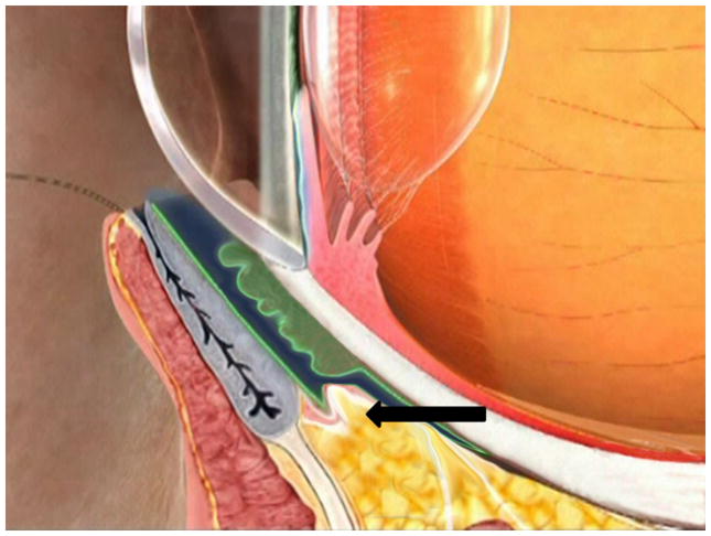
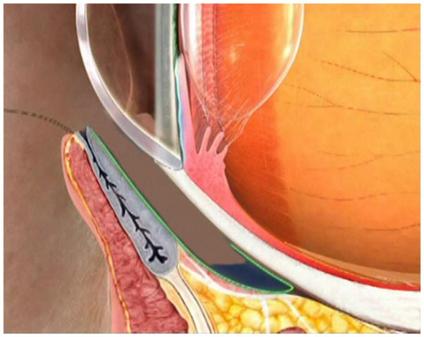
FIGURE 1. Schematic Demonstration of Fornix Tear Reservoir Obliterated by CCh and Restored by Fornix Reconstruction With Conjunctival Recession and AMT.
Under the normal circumstance, the fornix tear reservoir depicted in dark blue is responsible for delivering tear fluids to the tear meniscus by blinking (A, B). In CCh, the fornix tear reservoir is obliterated by loose and wrinkled conjunctiva (C, green) and prolapse fat (C, arrow) due to degenerated Tenon’s capsule. This results in low basal wettings which mimick ATD dry eye. Restoration of the fornix tear reservoir is achieved by fornix deepening reconstruction, resulting in disappearance of ATD dry eye secondary to CCh (i.e., converted to a normal state). Consequently, fornix deepening reconstruction also helps identify ATD dry eye that is independent of CCh (D).


