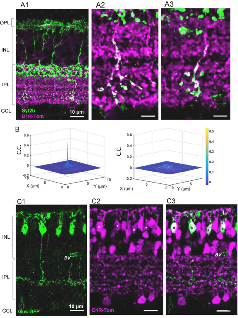
Figure 6.
Both type 6 and 7 ON bipolar cells colocalized with tdTomato fluorescence. (A1) Syt2b labeled axon terminals of type 6 cells and the entire structure of type 2 OFF bipolar cells (dendrites-somas-axons). Only axon terminals of type 6 cells colocalized with tdTomato. (A2-A3) Magnified views of type 6 axon terminals with tdTomato fluorescence in a single digital section (0.3 µm thick). (B) (left) 2D cross correlation coefficient showed that tdTomato fluorescence colocalized with syt2b-stained type 6 axon terminals (n=20 fields). (right) A 90° rotation of the green channel resulted in no correlation. (C1-C3) Double transgenic mice with Gus-GFP(C1) and Drd1a-tdTomato (C2) revealed that type 7 ON bipolar cells colocalized with tdTomato (C3). * indicates double-labeled cells, BV, blood vessel.

