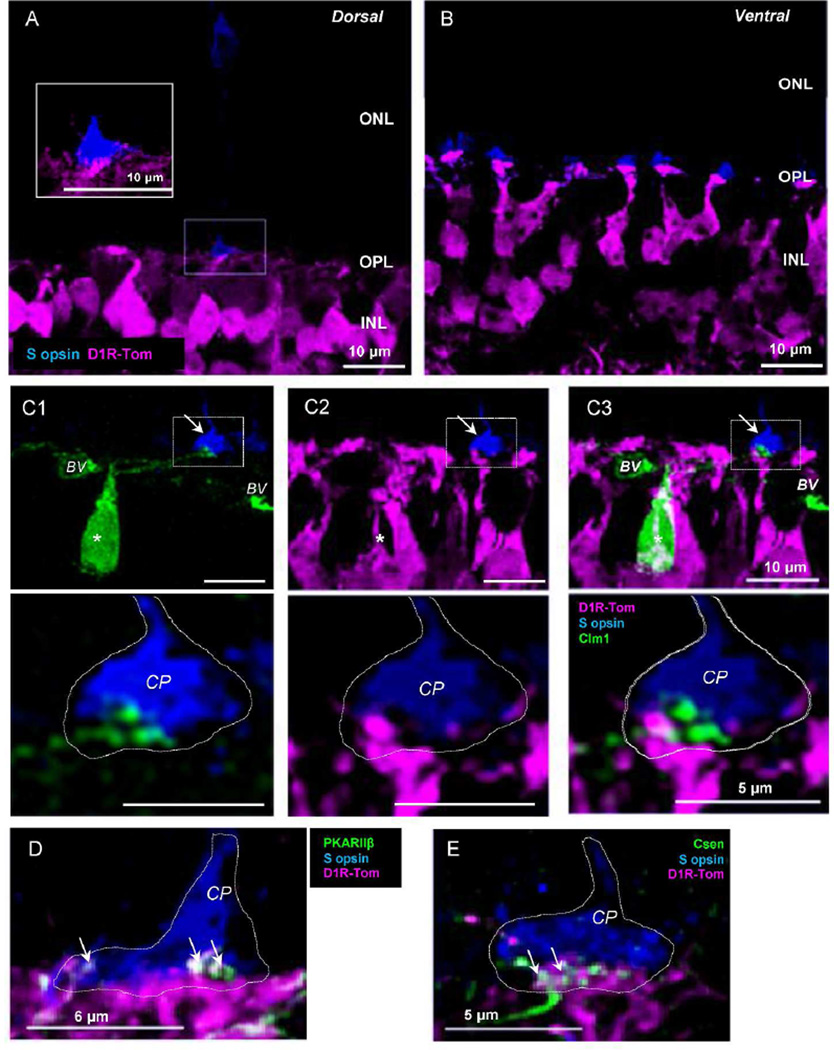
Figure 7.
Genuine S-opsin-expressing cells and tdTomato processes. (A) In the dorsal retina, S-cone terminals were occasionally observed, indicating that they were genuine S-cones. tdTomato fluorescent processes were attached to the terminal (see inset). (B) In the ventral retina, S-opsin-expressing cells were frequently observed, indicating that they were mixed cones with S-opsin and M-opsin. (C1 –C3) Double transgenic mice with tdTomato and Clm-1 revealed that type 9 cells (*) did not express tdTomato. Type 9 and tdTomato positive processes associated with S-cone pedicle were not colocalized (arrow and magnified image seen in bottom row). (D) tdTomato processes near Scones expressed PKARIIβ which is a type 3b OFF bipolar cell marker. (E) tdTomato processes attached to a genuine S-cone terminal were also partially colocalized with type 4 OFF bipolar cell dendrites (Csen). CP, cone pedicle. BV, blood vessel; CP, cone pedicle.

