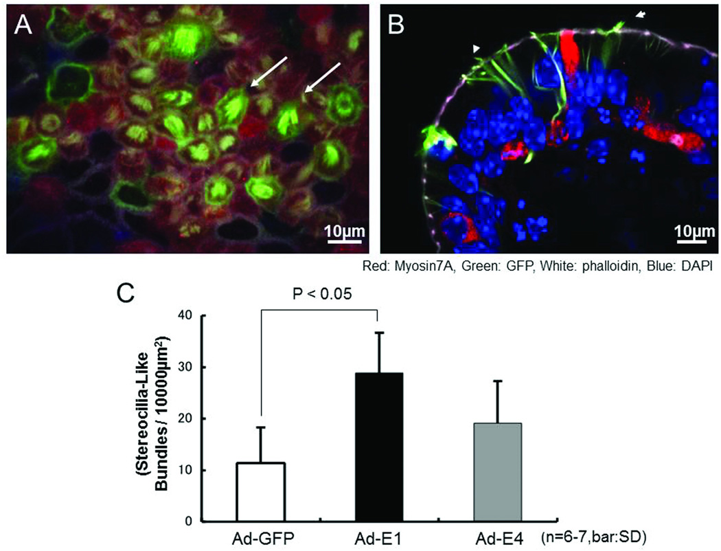
Figure 5.
Hair bundle-like structures on Ad-E1 transduced explants. A: Whole mount utricular explant. Many myosin7A-positive cells with hair-bundle-like structures on their apical surfaces are observed (e.g. arrows). B: Sectioned explant. Myosin7A-positive cell bodies are observed in the supporting cell layer. Hair-bundle-like structures protrude from extensions of these cells that reach the apical surface of the epithelium (e.g. arrowhead). Red: Myosin7A; Green: EGFP; Blue: DAPI. C: Number of hair-bundle-like structures observed after adenoviral transduction. In Ad-GFP transduced explants, a few damaged stereociliary bundles remained. However, in Ad-E1 transduced explants, there are many more immature hair bundles. In Ad-E4 transduced explants, some filamentous cilia-like structures are observed. The number of bundle-like structures in Ad-E1 transduced explants was significantly greater than in control (Ad-GFP) explants than in Ad-GFP transduced explants (p < .05, 6–7 explants per group)..

