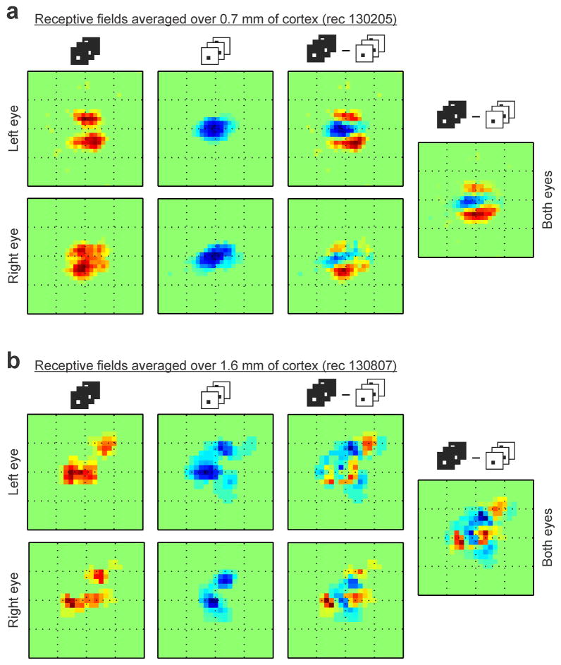
Extended Data Figure 2. ON/OFF domains are matched across eyes.
a, Integrating the ON/OFF receptive fields over 0.7 mm of horizontal cortical distance reveals ON and OFF receptive field subregions that are segregated in visual space and well matched between eyes. Notice the excellent binocular match of the receptive field subregions measured with light spots (left, two subregions displaced vertically in both eyes), and dark spots (middle left, one central subregion in both eyes). The ON-OFF receptive field difference also shows and excellent binocular match (middle right), therefore, the ON-OFF segregation can still be seen after combining the receptive fields of the two eyes (right). b, Integrating the ON/OFF receptive fields over a much longer distance (1.6 mm of cortex, different horizontal penetration) still reveals separate receptive field subregions with excellent binocular match. The 1.6-mm-average receptive-fields of the left and right eyes have both two ON subregions that are displaced diagonally and retinotopically matched (left). They also have two OFF subregions that are also displaced diagonally and retinotopically matched between the two eyes (middle left). A hint of the ON subregions can still be seen in the ON-OFF receptive field difference (middle right) and receptive field of both eyes combined (right), even if the receptive fields were averaged over 1.6 mm of cortex. Each square box framing a receptive field has a side of 16.2 degrees.

