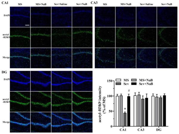
Fig. 5.
Effects of neonatal exposure to sevoflurane and sodium butyrate (NaB) treatment on the intensity of acetyl-H3K9 in hippocampal subfields. Representative images of the hippocampal CA1, CA3, and dentate gyrus (DG) areas of the rats from four groups: 4',6-diamidino-2-phenylindole (DAPI), acetyl-H3K9, and an overlay of DAPI and acetyl-H3K9 images. The intensity of acetyl-H3K9 in hippocampal CA1 (*P < 0.001 vs. the MS group; #P < 0.001 vs. the Sev group), CA3, and DG areas. The intensity of acetyl-H3K9 in the group was taken as 100%. n = 6 per treatment group. Scale bar = 50 μm. MS: maternal separation; Sev: sevoflurane.

