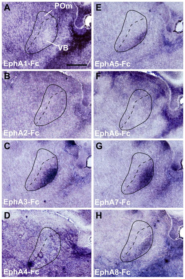
Figure 2. Patterns of EphA receptor binding in the VB and POm thalamic nuclei in the perinatal brain.
(A–H) Binding patterns of Fc-tagged EphA receptors (EphA1 – EphA8) within the VB and POm at P4. Note that the labeling by EphA3-Fc, A5-Fc, A7-Fc and A8-Fc binding shows a distinct gradient in the VB with the strongest labeling at the ventro-lateral region (C, E, G, H), whereas EphA1-Fc, A2-Fc, EphA4-Fc, and EphA6-Fc binding in the VB (A, B, D, F) does not. Scale bar = 500 μm.

