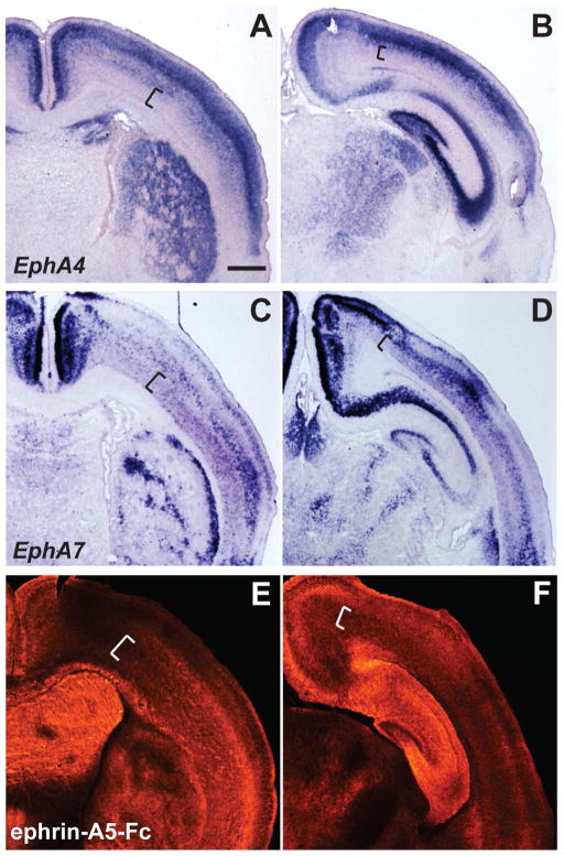
Figure 4. Distinct expression patterns of EphA4 and EphA7, in comparison to ephrin-A5 binding in the early postnatal brain.
(A–D) In situ hybridization of EphA4 (A and B) and EphA7 (C and D) in anterior (A, C) and posterior (B, D) sections of the P4 mouse brain. In both anterior and posterior sections, EphA4 is observed predominantly in upper cortical layers, with lower expression in deeper layers (A and B, brackets). In contrast, EphA7 labeling is evident in distinct gradients within deeper layers, including layer VI (C and D, brackets). Both EphA4 and EphA7 have distinct expression patterns in the striatum (A and C) and thalamus (B and D). (E and F) Binding patterns of ephrin-A5-Fc at anterior (E) and posterior (F) levels. The relative labeling intensity of ephrin-A5-Fc binding displays an overall similarity to EphA7 expression, including the gradients in layer VI (see brackets), striatum, and thalamus. Scale bar = 500 μm.

