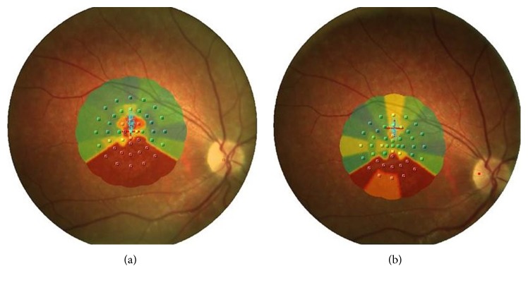
Figure 1.
(a) Interpolated microperimetric map of RE before rehabilitation at zero time in which we see the image of foveal atrophy that does not allow a good fixation of the target (red cross). (b) Interpolated microperimetric map at the end of rehabilitation training after 12 months in which an improvement of fixation stability through the identification of TRL was achieved.

