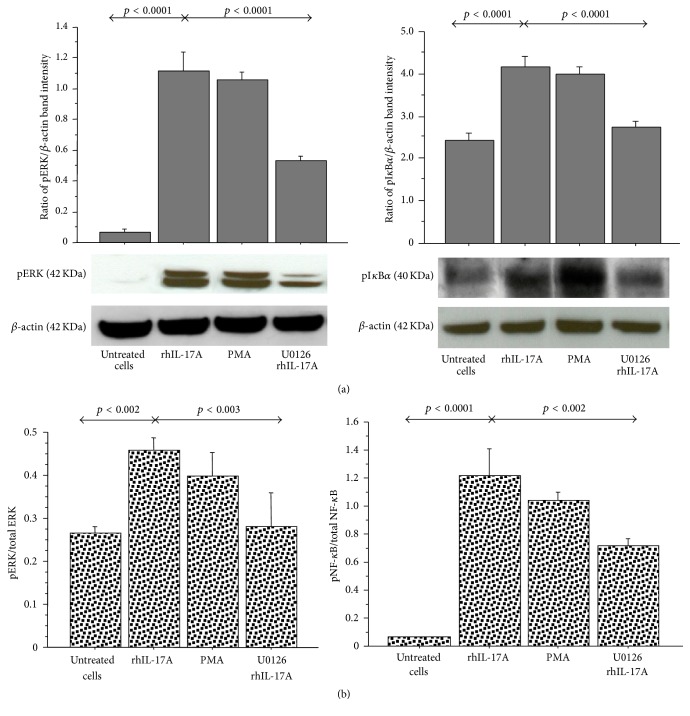
Figure 1.
Effect of U0126 inhibitor on ERK and IκBα phosphorylation in RPMI 2650 cells stimulated with rhIL-17A. The cells were stimulated with rhIL-17A (20 ng/mL) or PMA (50 ng/mL) for 30 min in absence or presence of U0126 (25 μM). (a) pERK and pIκBα protein expression were evaluated in the cell lysates by western blot. The results were expressed as ratio of band intensity and β-actin of 3 separate experiments. Representative gel images of pERK, pIκBα, and β-actin are shown. (b) The activation of ERK1/2 and NF-κB for each experimental condition was tested for the pERK1/2/total ERK1/2 ratio and for the pNF-κB/total NF-κB, respectively, by ELISA and normalized for protein content. ANOVA with Fisher's test correction was used for the analysis of the data. p < 0.05 was statistically significant.

