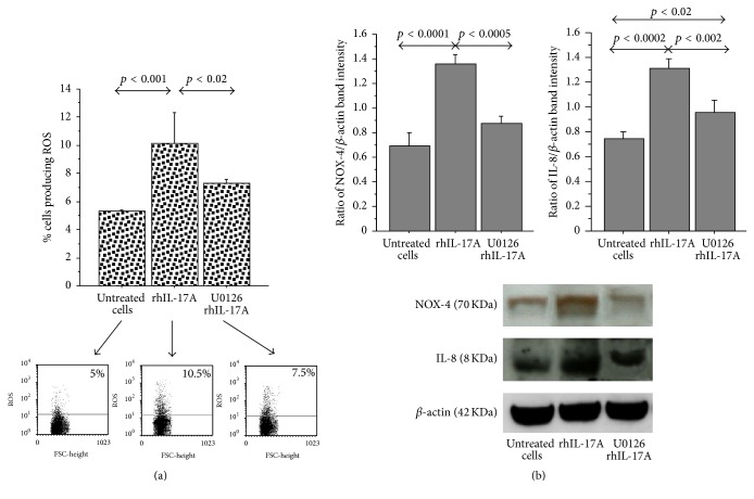
Figure 2.
Effect of U0126 inhibitor in RPMI 2650 cells stimulated with rhIL-17A. (a) The cells were stimulated with rhIL-17A (20 ng/mL) for 6 hrs in absence or presence of U0126 (25 μM). ROS production was evaluated in the cells by flow cytometry. The bars represent the mean ± SD of 3 separate experiments. Representative flow cytometry are shown; (b) the cells were stimulated with rhIL-17A (20 ng/mL) for 18 hrs in absence or presence of U0126 (25 μM). NOX-4 and IL-8 protein expression were evaluated in the cell lysates by western blot. The results were expressed as ratio of band intensity and β-actin of 3 separate experiments. Representative western blot is shown. ANOVA with Fisher's test correction was used for the analysis of the data. p < 0.05 was statistically significant.

